Cardiac (Please use Links above to Navigate This Section)
Anatomy
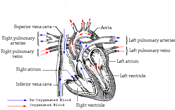
The heart could be described as being 2 pumps. 1 pump (right side) sends blood to your lungs to be oxygenated and to remove waste products (CO2) and the other pump (left side) sends the blood around the systemic circulation to oxygenate all the cells in the body. The heart weighs between 7 and 15 ounces (200 to 425 grams) and is a little larger than the size of your fist, it is located between your lungs in the middle of your chest, behind and slightly to the left of your breastbone (sternum).
The heart has 4 chambers. Two upper chambers are called the left and right atria, and the two lower chambers are called the left and right ventricles. The septum (a wall of muscle) separates the left and right atria and the left and right ventricles. The left ventricle is known as the largest and strongest chamber in your heart with enough force to push blood through the aortic valve and into your body.
The heart chambers have valves which assist in the transport of blood flow through the heart, These are:
- The tricuspid valve regulates blood flow between the right atrium and right ventricle.
- The pulmonary valve controls blood flow from the right ventricle into the pulmonary arteries, which carry deoxygenated blood to your lungs to oxygenated.
- The mitral,or bicuspid valve lets oxygenated blood from your lungs pass from the left atrium into the left ventricle.
- The aortic valve opens the way for oxygenated blood to pass from the left ventricle into the aorta, your body's largest artery, from here the blood is distributed to whole of your body.

Electrical System
The electrical system in your heart controls the speed of your heartbeat. Your heart has three main components to the system, these consist of:
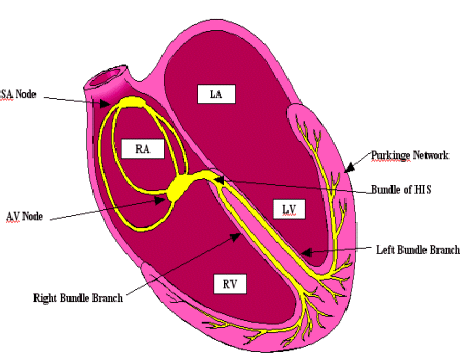
- S-A node (sinoatrial node)
- A-V node (atrioventricular node)
- Purkinje system
The S-A node, also called the "natural pacemaker", of your heart because it controls your heart rate. The S-A node is made of specialised cells located in the right atrium of the heart. The S-A node creates the electricity that makes your heart beat. The S-A node normally produces 60-100 electrical signals per minute — this is your heart rate.
The A-V node is a bundle of cells between the atria and ventricles. The electrical signals generated by the S-A node are "caught" and held for milliseconds before being sent onto the bundle of HIS (HIS Purkinje system).
HIS purkinje system is in your heart's ventricles. Electricity travels through the His-purkinje system to make your ventricles contract. The electricity from the A-V node hits the bundle of HIS before being directed into the right and left bundle branches and finally into the purkinje fibres that are located in the cardiac muscle. This stimulates the ventricles to contract.

The Electrical Pathway
- STEP 1. The S-A node creates an electrical signal
- STEP 2. The electrical signal follows natural electrical pathways through both atria. The movement of electricity stimulates the atria to contract, which pushes blood into the ventricles.
- STEP 3. The electrical signal reaches the A-V node. There, the signal pauses to give the ventricles time to fill with blood
- STEP 4. The electrical signal spreads through the His-purkinje system. The movement of electricity causes the ventricles to contract and push blood out to your lungs and body.
The name for the steps above is known as the cardiac cycle which lasts for 0.8 seconds:
- Atrial systole = 0.1 second
- Ventricular systole = 0.3 seconds
- Diastole = 0.4 seconds
- Systole refers to the contraction of the cardiac muscle
- Diastole refers to the relaxation of the cardiac muscle
Nervous Control of the Heart
Although the S-A node sets the basic rhythm of the heart, the rate and strength of its beating can be modified by two auxiliary control centres located in the medulla oblongata of the brain.
- One sends nerve impulses down accelerator nerves.
- The other sends nerve impulses down a pair of vagus nerves
Accelerator Nerves
The accelerator nerves are part of the sympathetic branch of the autonomic nervous system. They increase the rate and strength of the heartbeat and thus increase the flow of blood. Their activation usually arises from some stress such as fear or exertion. The heartbeat may increase to 180 beats per minute. The strength of contraction increases as well so the amount of blood pumped may increase to as much as 25-30 litres/minute.
Vagus Nerve
The vagus nerves are part of the parasympathetic branch of the autonomic nervous system. They, too, run from the medulla oblongata to the heart. Their activity slows the heartbeat.
Pressure
receptors in the aorta and carotid arteries send impulses to the medulla which relays these impulses back by way of the vagus nerves to the heart. Heartbeat and blood pressure diminish.